
SEARCH
Wellgen's Next Gen Digital Cytology System captures eosinophils in striking cellular detail.
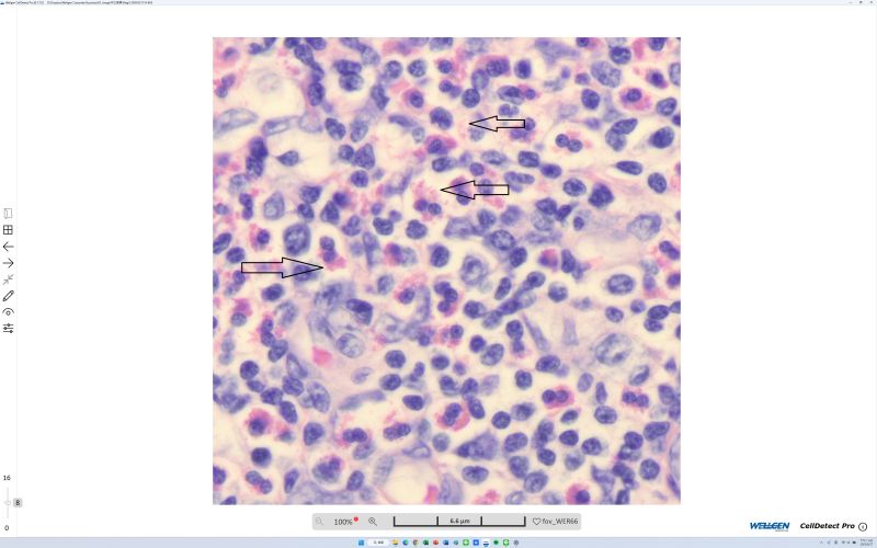
Ever seen eosinophils under a bright-field microscope? Take a look at this—can you see the reddish particles? Eosinophils (also known as eosinophilic leukocytes) are characterized by their association with allergic reactions, parasitic infections, and certain tumors. Their numbers typically rise when the body is stimulated by these conditions, and when excessively elevated, the substances they release can trigger tissue inflammation and cell damage, potentially leading to symptoms such as asthma or inflammation in organs like the lungs and esophagus.
This image was captured by Wellgen Medical's Next Gen Digital Cytology System, the most advanced automated microscope to use tomographic scanning technology for cytology. The system enables observation of cytology smears at routine 450x, 900x, and even 1,300x magnification—without the need for an oil lens or complex scanning electron microscopy (SEM).
By eliminating the complexity traditionally required for high-magnification cytology, Wellgen makes cellular-level detail more accessible for clinical and research use alike. Interested in seeing your own cells beyond 1,000x under a bright-field microscope? Contact Wellgen to learn more—another step forward in the company's mission of "AI-Assisted Diagnosis."
依據歐盟施行的個人資料保護法,我們致力於保護您的個人資料並提供您對個人資料的掌握。
按一下「全部接受」,代表您允許我們置放 Cookie 來提升您在本網站上的使用體驗、協助我們分析網站效能和使用狀況,以及讓我們投放相關聯的行銷內容。您可以在下方管理 Cookie 設定。 按一下「同意」即代表您同意採用目前的設定,更多資訊請瀏覽 隱私權聲明。